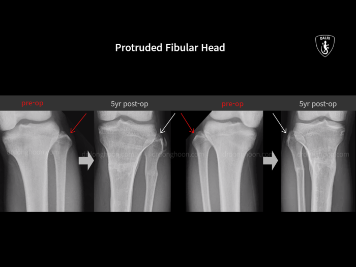
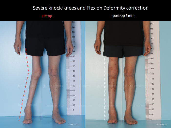
Femoral lengthening
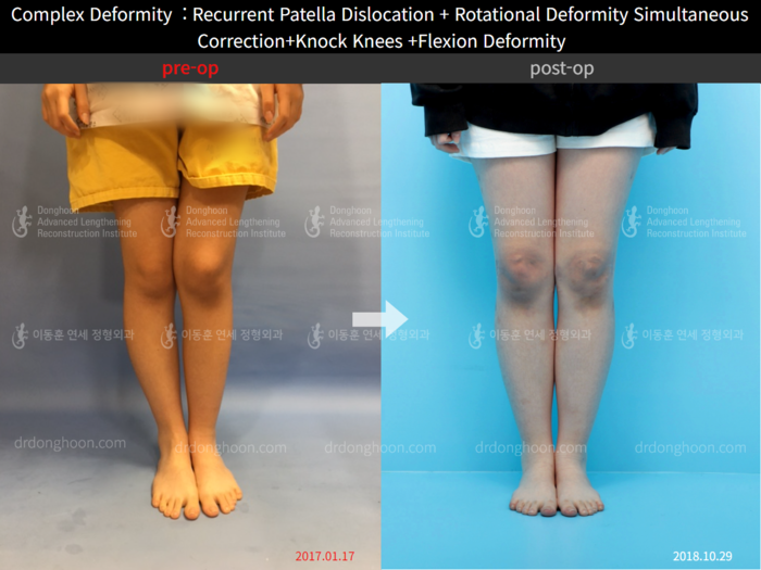
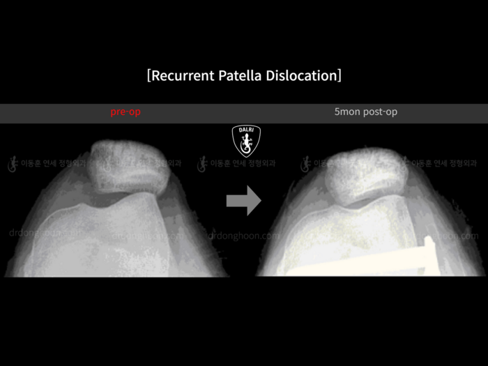
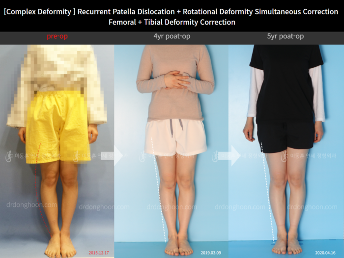
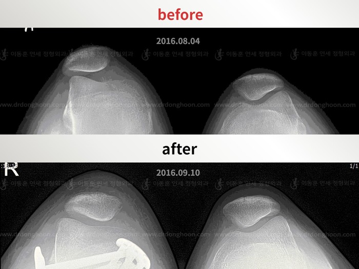
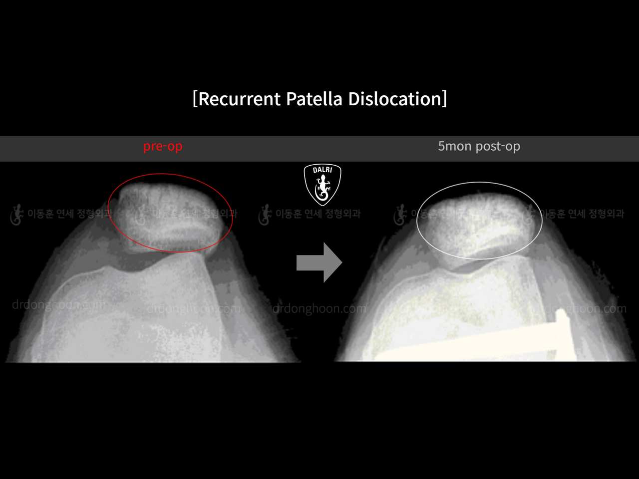
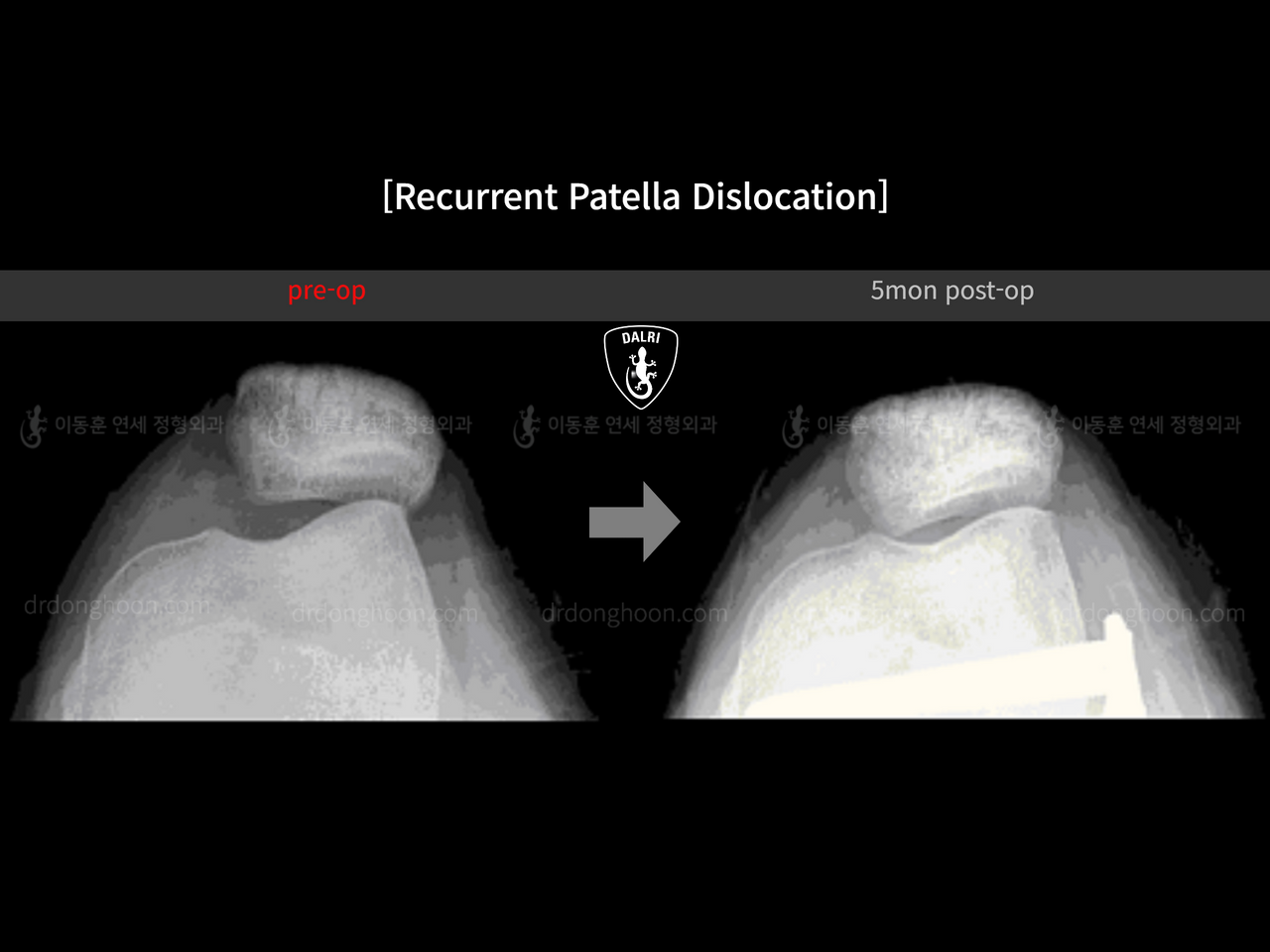
A recurrent patella dislocation case at the Dr. Donghoon institute (DALRI), Korea (5 months post-op), with 8 before and after radiographs.
- Category
- Recurrent patella dislocation
- Gained
- —
- Height
- —
- Follow-up
- 5 months
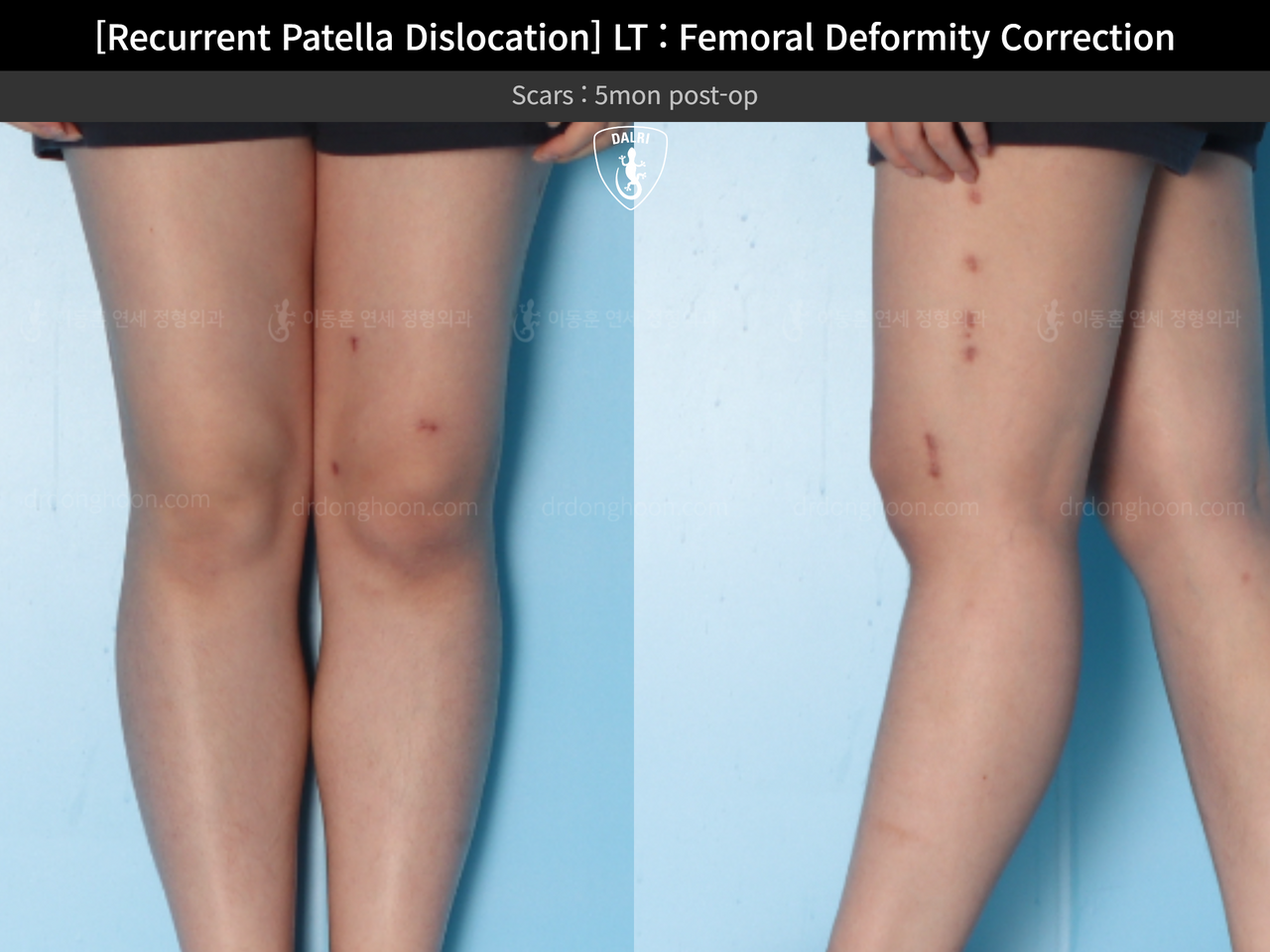
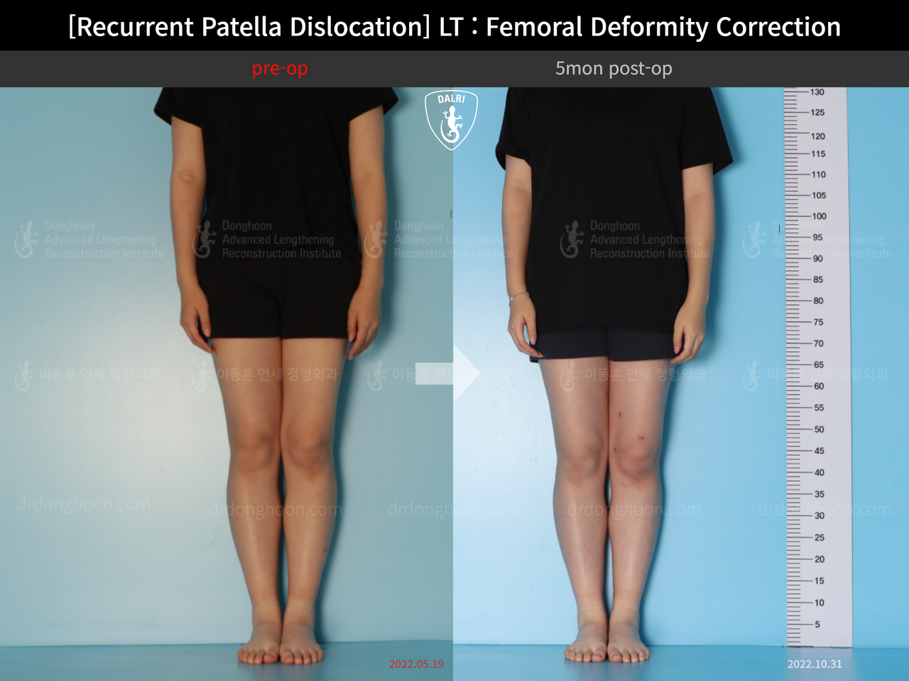
Drag the handle to compare before and after (arrow keys work too). Before and after recurrent patella dislocation, case 13337596, image 2, Dr. Donghoon institute (DALRI)
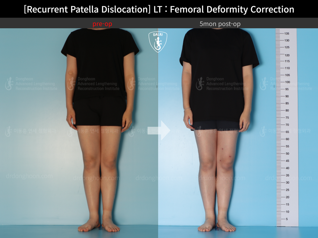
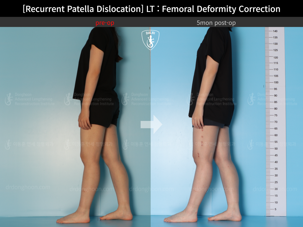
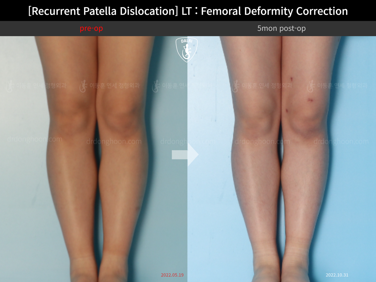
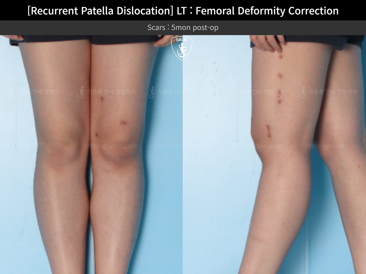
About this case
This patient was treated in the congenital & paediatric conditions group (recurrent patella dislocation) at the Dr. Donghoon institute (DALRI) in Seoul.
This is an alignment or limb-length correction, so the source record carries no centimetre gain; the result is read from the mechanical axis on the radiographs instead.
The series on this page contains 8 images, in the order the hospital published them.
Technique and segment
The source record does not name the technique used here. The choice between an external frame, an internal nail, or a hybrid depends on the segment, the target length, and each patient's soft-tissue condition.
The femur is wrapped in well-vascularised muscle, so the new bone usually consolidates faster than in the tibia and tolerates a larger gain per segment, at the cost of watching for knee stiffness and quadriceps weakness.
Timeline and how to read the radiographs
In deformity-correction cases the distraction phase is shorter than in height cases, because the goal is realignment rather than distance gained.
This series was taken 5 months after surgery. The regenerate is not yet fully opaque, which is the expected appearance at this stage.
Daily physiotherapy through the distraction phase is the single strongest predictor of the result: patients who stop stretching run into joint stiffness far more often than into problems with the bone itself.
All images are published by the Dr. Donghoon institute (DALRI) and shown with permission. Individual results vary; these cases are not a guarantee of outcome.